
Microscope Accessories and Replacement Parts
General accessories and parts for use with microscopes. Products include lens oil, lens cleaner, slide dispensers, microscope covers, vessel holders, light cubes, and flow cells.
Many parts of your microscope can be updated or replaced including microscope lamps, lenses, and eyepieces. Replacement lamps, light sources, and power cords can extend the working lives of your microscopy equipment. New or replacement objectives and lenses can help increase the total magnification available with your microscope.
Add greater magnification or exchange your eyepiece for one with a reticule to more easily estimate the sizes of the items being viewed.
Replace or update your mechanical stage or clamps or add cell culture vessel adapters to facilitate specimen positioning.
Condensers can also be replaced, and conversion kits are available to add phase contrast, polarization, fluorescence, or other capabilities to your current system.
Some microscopes can be adapted for use with still or video cameras. Look for cameras and adapters designed for your specific microscope brand. Film and documentation-related products may also be available to support your record-keeping.
Temporary, semi-permanent, and more long-term slide storage options include:
- Slide racks that securely hold slides during staining procedures
- Folding fiberboard or plastic slide holders with horizontal indentations that keep microscope slides in place
- Slide storage boxes with vertical dividers; made from fiberboard, plastic, and wood and available in multiple sizes
- Slide holders and mailers for transporting slides safely
- Slide storage systems and cabinets for large quantities of slides or archival storage


Electron Microscopy Sciences EMS Platinum Aperture with 15 µm hole, 0.53 mm thick
Platinum apertures are easily cleaned by either an aperture flamer or over a bunsen burner in the open air. We maintain a wide variety of Platinum Drilled Apertures in stock with different configurations suiting all of your needs. Nominal hole diameter: +/- 1 micron. Disc diameter: +/- 10 microns. Disc thickness: +/- 10 microns
Size: 15 µm
O.D.: 2 mm
Thickness: 0.53 mm

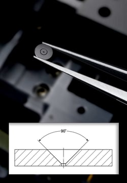
Electron Microscopy Sciences EMS Gold Aperture with 200 µm hole, 3 mm O.D., 0.12 mm thick
Advanced techniques of thin metal film application permits the apertures to retain a clean hole edge during prolonged beam exposure. Ultra-thin, half-micron design discourages contaminant accumulation and stabilizes astigmatic conditions to provide longer life. Conventional mechanical cleaning involving costly down-time is no longer necessary. Because the foil is ultra thin, care must be taken when handling the gold foil apertures.
Size: 200 µm
O.D.: 3 mm
Thickness: 0.12 mm

Electron Microscopy Sciences K-kits Liquid TEM Kit, 2.0 µm Gap
An Innovative Enclosed Specimen Holder for Liquid TEM. K-kits are sample holders designed to facilitate convenient TEM observation of liquid samples, allowing nanoobjects, aggregates, and agglomerates (NOAAs) in liquid samples to be characterized. With vacuum compatible sealing of liquids in electron-transmitting thickness, K-kits are micro reaction chambers for countless experiments in materials, chemical, and biological research.
Gap: 2.0 µm

Electron Microscopy Sciences TEM Screen Recoating Phillips Main Large
For optimum viewing, fluorescent screens should be recoated regularly. Viewing screens are easily damaged by the electron beam, especially at high accelerating voltages where knock-on damage can be severe. A proprietary process where 1 to 2 micron particles of phosphor are deposited on the screen and produce the highest possible resolution.

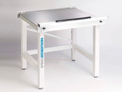
Electron Microscopy Sciences AMD-FH Microscope Workstation, 75 x 75 cm, Ferromagnetic SS Surface
These isolation tables support most of the larger microscopes and their support equipment. The air isolation platform effectively removes most removes most vibrations. The rigid frame has levelling feet which ensure the table is in excellent contact with the floor. The damped rolling diaphragm isolators have a low natural frequency of 2Hz which effectively isolates out vibrations above 6Hz, well below the frequencies at which typical building vibrations begin to occur. Full Active table with self-levelling and platform guards.
Size: 75 x 75 cm
Surface: Ferromagnetic SS
Type: Active Air Self Leveling

Electron Microscopy Sciences Gilder Thin Bar Hexagonal 600 mesh Copper Grid
Very fine mesh grids have up to 2,000 lines/inch. There is an increasing need in TEM for support films, routinely carbon, as thin as 1.5 - 2.0 nm. The pitch (the distance from the center of one bar to the center of the next bar) dimension in all grids remains constant, allowing use as a lower magnification calibration aid.
Apart from one side being shiny, the other matt, there is a large asymmetrical mark in the rim which gives the identification of which side the specimen is on. Grids are 3.05 mm overall diameter; 2.05 mm mesh area diameter. With 5 µm bars more of the specimen can be viewed then in standard grids.
Grid: Copper
Pitch: 42 µm
Hole: 37 µm
Bar: 5 µm
:

Electron Microscopy Sciences Indexed Grid Graticle NE34A, 19 mm, 1 x 1 mm with 1.0 mm pitch
Useful for particle counting, particularly when reference is needed between workers. Also, they are useful for area of specimen determinations. Indexed grid marked 1-10 and A-J.
Size: 19 mm
Divisions: 1 x 1 mm with 1.0 mm pitch
Surface: Chrome image

Electron Microscopy Sciences Gilder Hexagonal 400 mesh Gold Grid
A reliable support specimen grid source. Features: well-defined grid bars, maximum open area, and a matt/shiny side. Each grid is individually inspected. The copper grids with palladium plating offers better grid strength and avoids tarnishing. 3.05 mm diameter, 0.7 mil (18um) thickness.
Grid: Gold
Pitch: 62 µm
Hole: 37 µm
Bar: 25 µm

Electron Microscopy Sciences V-1 Quality, 50 x 75 mm, 0.15- 0.21 mm thick
EMS Mica Sheets offer a clean surface for EM applications, carbon filming and particle spraying, as well as for AFM applications. There are two types of mica: muscovite and phlogopite. Generally, one differs from the other by color (Muscovite is Ruby, Green or White; Phlogopite is Amber, Yellow, or Silver). The maximum operating temperature for Muscovite is about 500-600 degree C and for Phlogopite is about 800-900 degree C. Our line consists of High Quality Muscovite Mica. This mica peels off very thin up to 0.0001" uniform layers, exposing "virgin" mica upon splitting.
Muscovite, potash type mica, sometimes known as granitic mica, is the best of all micas in dielectric strength, perfection of cleavage, and transparency. It has the chemical formula: H2KAI3(Si04)3. Clear. Hard, of uniform color, nearly flat, free of all stains, foreign inclusion, cracks, and other similar defects.
Quality: V-1
Size: 50 x 75 mm
Thickness: 0.15- 0.21 mm
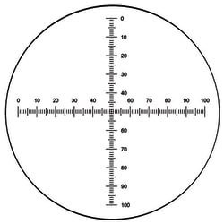
Electron Microscopy Sciences Crossed Scale Microscope Graticle NE17, 21 mm, 10 mm with 0.1 mm subdivisions
Used as a horizontal and a vertical scale, they are especially useful when interested in measurement in different axis. Crossed micrometer scale.
Size: 21 mm
Divisions: 10 mm with 0.1 mm subdivisions
Surface: Chrome image

Electron Microscopy Sciences Tripod Polisher PrecISIon Locking Micrometer
Special larger diameter locking rings for easy locking and are graduated in one micron increments with 13mm travel. Non-coating Micrometers not recommended. - For use with Tripod Polisher.
Electron Microscopy Sciences V5 Quality, 25 x 25 mm, 0.15- 0.21 mm thick
EMS Mica Sheets offer a clean surface for EM applications, carbon filming & particle spraying, as well as for AFM applications. There are 2 types of mica: muscovite & phlogopite. Generally, one differs from the other by color (Muscovite is Ruby, Green or White; Phlogopite is Amber, Yellow, or Silver). The maxoperating temp.for Muscovite is about 500-600 degree C & for Phlogopite is about 800-900 degree C. Our line consists of High Quality Muscovite Mica. This mica peels off very thin up to 0.0001" uniform layers, exposing "virgin" mica upon splitting.
Muscovite, potash type mica, sometimes known as granitic mica, is the best of all micas in dielectric strength, perfection of cleavage, & transparency. It has the chemical formula: H2KAI3(Si04)3. Stained A Quality. Hard, med. air inclusions uniformly distributed in usable area; slight green vegetable stains, med. waviness, & heavy waves if specified.
Quality: V5
Size: 25 x 25 mm
Thickness: 0.15- 0.21 mm

Electron Microscopy Sciences TEM Screen Recoating Phillips Alignment
For optimum viewing, fluorescent screens should be recoated regularly. Viewing screens are easily damaged by the electron beam, especially at high accelerating voltages where knock-on damage can be severe. A proprietary process where 1 to 2 micron particles of phosphor are deposited on the screen and produce the highest possible resolution.
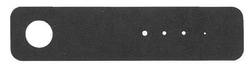
Electron Microscopy Sciences EMS Strip Aperture Hitachi, 10-30-50-70 µm holes, 45.0 x 4.0 x 0.10 mm
Strip apertures made from Platinum-Iridium (95:5).
Holes: 10-30-50-70 µm
Dimensions: 45.0 x 4.0 x 0.10 mm
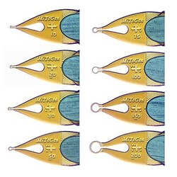
Electron Microscopy Sciences MicroMounts™ Crystal Tool with 200 µm Aperture and 19 mm pin
MicroMounts are the high performance tools for manipulating and mounting protein crystals, virus crystals, and small molecule/inorganic crystals, and other small, fragile samples. They are ideal for both conventional and high-throughput X-ray crystallography applications.
Each consist of a thin (7.5um thick) micro-fabricated polyimide film, attached to a solid non-magnetic stainless steel pin. Film is wrapped around the steel pin and it provides excellent stiffness and convenient scoop-like action in retrieving and handling crystals.
Aperture: 19 mm pin